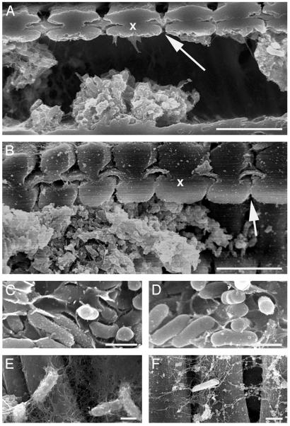
Fig. 6.
Scanning electron micrographs of cells and associated exopolysaccharide from the wild-type strain Temecula (A, C, and E), and the rpfF mutant KLN61 (B, D, and F) in the xylem of grapevine petioles from symptomatic leaves. (A and B) Wild-type and rpfF communities were similar (x, two adjacent xylem vessel walls; arrow, bordered pit with pit membrane). Cells in crowded vessels were embedded in a matrix (C and D) whereas individual cells were covered with strands of potentially the same material (E and F).

