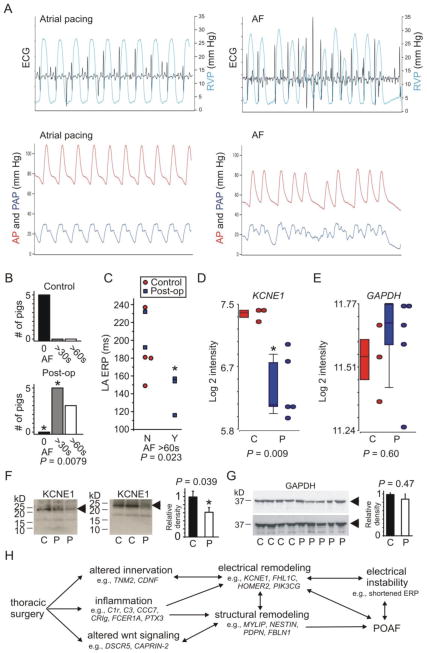
Figure 1. Atrial KCNE1 downregulation in porcine postoperative atrial fibrillation.
A. Representative tracings of ECG, right ventricular pressure (RVP), systemic arterial pressure (AP), and pulmonary arterial pressure (PAP) during atrial pacing at a rate of 80/min and following induction of AF
B. Incidence of sustained AF in Control and 3-days postoperative pigs. *P=0.0079 versus Controls
C. Left atrial effective refractory period duration versus incidence of AF sustained >60 s after 10 s rapid pacing (Y, yes versus N, no) plotted for control and 3-days postoperative pigs as indicated
D. Mean KCNE1 transcript intensity in LA tissue of Control (C) and postoperative (P) pigs; n = 3–5
E. Mean GAPDH transcript intensity in LA tissue of Control (C) and postoperative (P) pigs; n = 3–5
F. Left, exemplar western blots using anti-KCNE1 antibody to probe lysates from LA tissue of Control (C) and postoperative (P) pigs; right, band densitometry for blots as in left; n = 4–5, each performed in triplicate
G. Left, exemplar western blots using anti-GAPDH antibody to probe lysates from LA tissue of Control (C) and postoperative (P) pigs; right, band densitometry for blots as in left; n = 4–5, each performed in triplicate
H. Hypothetical framework of POAF etiology drawing from the findings herein and previous functional analyses of the genes listed.

