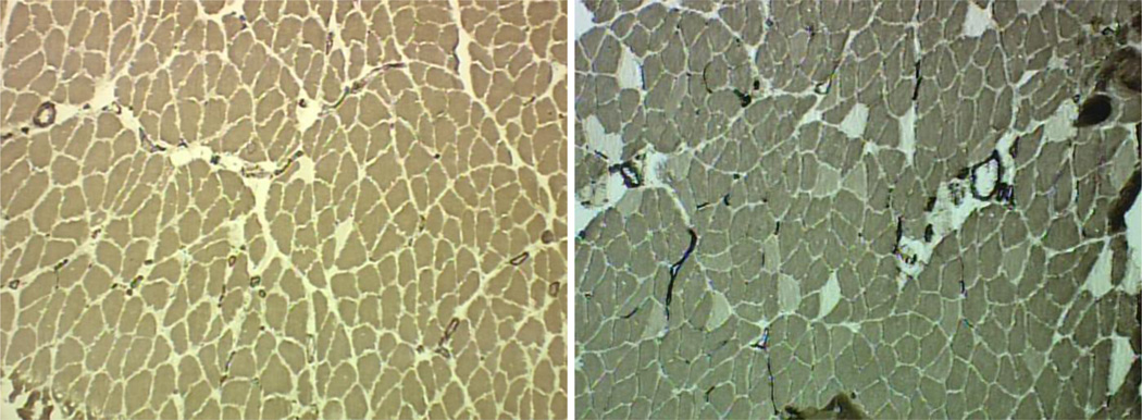
Figure 2.
Histochemically stained myofibers of aged (left panel) and young adult (right panel) soleus muscles. Darkest fibers are Type I, lightest fibers are Type IIA, and intermediate stained fibers are Type IIX. Note smaller fiber size and limited number of Type II fibers in the aged soleus. Original magnification of 100 x.

