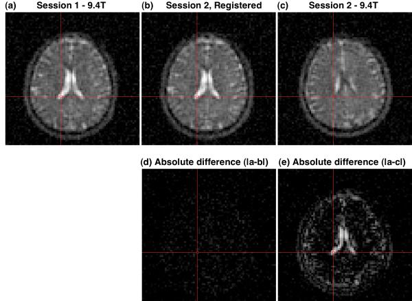
Figure 6.
Example of registration of 9.4 T sodium 23Na images (a,c) from different imaging sessions (1,2) of the same subject. The images from the session 1 (a) and session 2 (c) have different subject coordinate systems because of different head positions for the two sessions. After determining the alignment transform using AIR in the image domain, the images from session 2 were aligned to the images from session 1 by applying the transform in the k-space domain and then reconstructing these session 2 data in the session 1 subject coordinate system. The features of the resultant image (b) match those in session 1 (a) as demonstrated by the near zero absolute difference image (d). By comparison, the absolute difference image for the unaligned images (a and c) shows significant non-zero features (e). The absolute difference images (d and e) are shown on the same gray scale as (a and c).

