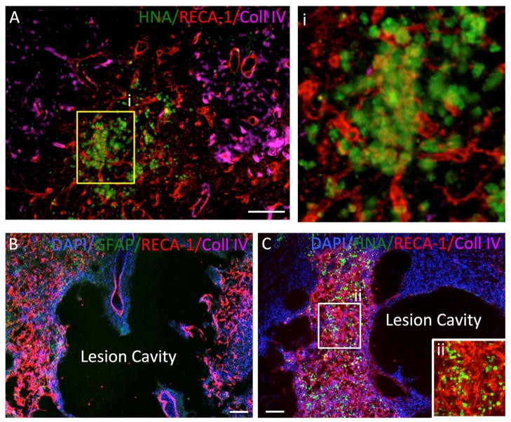
Figure 3. Implantation of hNSC into normal and stroke-damaged brain.
A. Human neural stem cells (HNA in green) injected into the normal rat striatum induce an angiogenic response (Reca-1 in red) at their site of injection with blood vessels penetrating the graft, but also individual endothelial cells being present (boxed area enlarged in i). Importantly these newly formed vessels do not yet contain a basement membrane as indicated by collagen IV (in pink). B. Stroke induces a dramatic angiogenic response in the peri-infarct area and this is associated with astrocytes (GFAP in green). C. A peri-lesion invasion of injected human hNSCs (HNA in green) indicates a co-localization with an area undergoing neovascularization. This neovascularization of the peri-infarct area is a gradual process and it is evident that other areas have not yet developed a dense vascular network. No ingrowth into the lesion cavity can be observed after injection of hNSCs. A higher magnification of the area undergoing angiogenesis indicates that implanted hNSCs are integrated within the mesh of newly forming blood vessels (ii).

