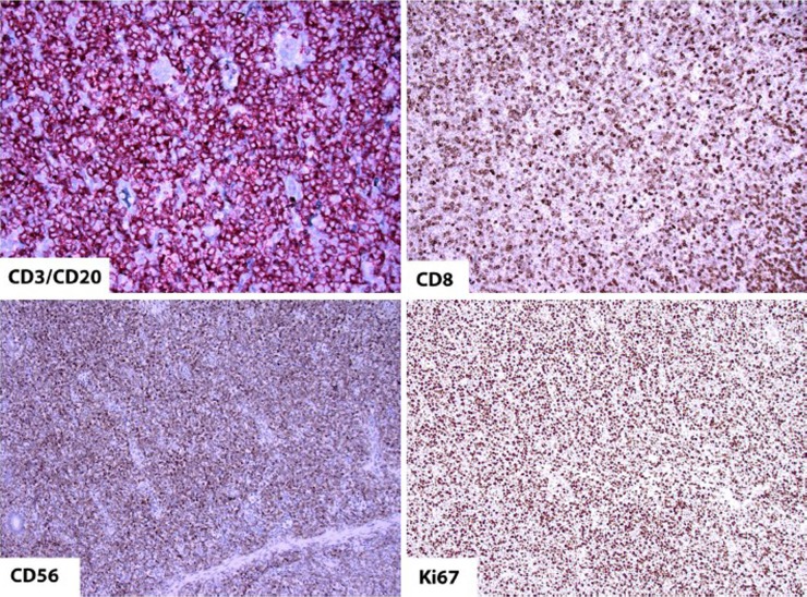
Figure 5.
The CD3/CD20 stain, a polymer cocktail consisting of anti-mouse/alkaline phosphatase and anti-rabbit/horseradish peroxidase demonstrates diffuse infiltration of CD3 positive neoplastic T cells (red), whereas, only rare CD20 positive B cells (brown) are present (top left, IHC, 100×). The tumor cells co-express CD8 (top right, IHC, 100×) and CD56 (bottom left, IHC, 100×) and show a high (~80-90%) proliferation index as evident by tumor cell nuclei staining for Ki-67 (bottom right, IHC, 100×)

