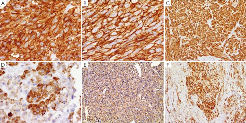
Figure 5.
Immunohistochemical features of GIST. A. Spindle cell GIST with strong and diffuse cytoplasmic staining of CD117 (c-kit) (×400); B. Spindel cell GIST with strong and diffuse membrane staining of CD34 (×400); C. Epithelioid cell GIST with strong cytoplasmic staining of CD117 (×100); D. Epithelioid cell GIST with patchy and heterogeneous staining of CD34 (×400); E. Epithelioid cell GIST with punctate staining of h-Caldesmon (×100); F. Epithelioid cell GIST with patchy mambrane staining of h-Caldesmon (×400)

