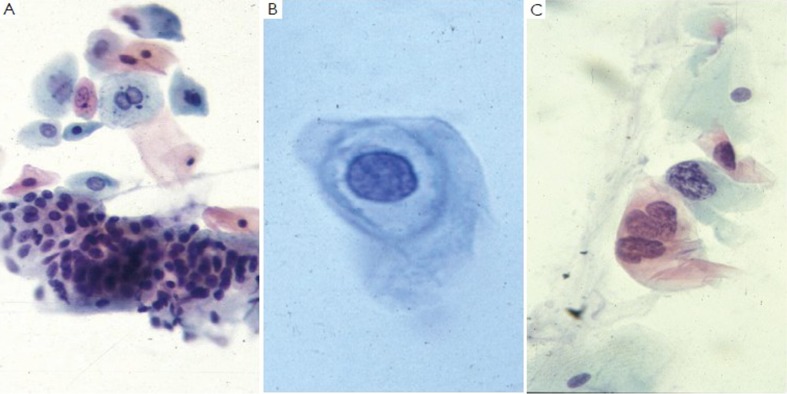
Figure 17.
A. normal anal Pap with intermediate and basal squamous cells and glandular cells (Pap stain, 400×); B. AIN1, showing a koilocyte with a prominent perinuclear cavity (Pap stain, 400×); C. AIN 3, displaying increased nuclear:cytoplasmic ratios and irregular hyperchromatic nuclei (Pap stain, 400×)

