Figure 5.

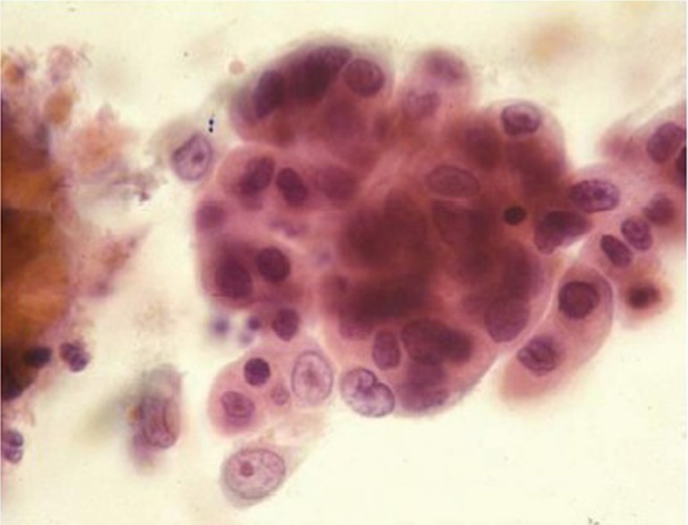
Esophageal adenocarcinoma with clusters of overlapping cells and single cells displaying delicate cytoplasm, enlarged irregular nuclei, prominent nucleoli, and necrotic background (Pap stain, 400×)

Esophageal adenocarcinoma with clusters of overlapping cells and single cells displaying delicate cytoplasm, enlarged irregular nuclei, prominent nucleoli, and necrotic background (Pap stain, 400×)