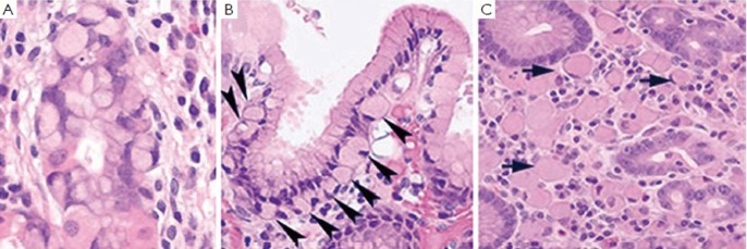
Figure 7.
A.In situ signet ring carcinoma cells confined within basement membrane; B. Pagetoid spread of signet ring cells (arrow heads) below the preserved surface epithelium; C. Focus of intramucosal signet ring cell carcinoma (arrows) in the lamina propria (all three photos are courtesy of Dr. Rebecca Fitzgerald)

