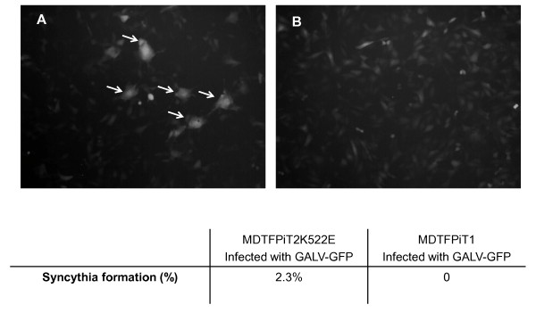
Figure 8.
Syncytia formation observed in MDTFPiT2K522E cells infected with GALV-GFP. MDTFPiT2K522E (A) or MDTFPiT1 cells (B) were exposed to GALV-GFP viruses. Four days after viral exposure, Several GFP positive syncytia were observed in MDTFPiT2K522E cells and the number of syncytia gradually increased over time. The experiments were performed three times and the representative images from cells infected with GALV-GFP seven days after viral exposure are shown. Syncytia were defined as those cells containing three or more nuclei. The total number of nuclei in syncythia cells was then counted. Five random fields were counted from each well of triplicate samples, using a 10x objective [11].

