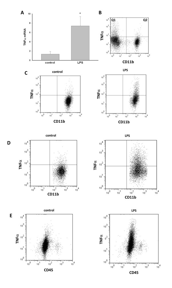
Figure 6.
Analysis of the microglial phenotype. We compared the phenotype of microglia isolated from the brains of control and LPS-treated mice. (A) qRT- PCR identified significantly upregulated levels of TNF-α mRNA in CD11b+ cells isolated from LPS-treated mice. TNF-α protein content in CD11b+ cells was compared (B, C) before and (D) after the immunomagnetic isolation procedure. (B) In brain tissue homogenates from control mice, only a small percentage of microglia contained TNF-α (Q2), but there was a CD11b negative cell population with high TNF-α content (Q1). (C) TNF-α in brain tissue homogenates gated on CD11b+ cells. (D) The analysis of TNF-α in isolated microglial cells showed that the frequency of TNF-α-positive cells increased similarly (by 6-fold) in CD11b+ cells before and after isolation. (E) TNF-α was produced predominantly by CD11b+/CD45low cells. The analysis of CD45 expression showed that there was not substantial infiltration of peripheral macrophages 20 hours after intraperitoneal injection of LPS, as we did not detect increased CD45 levels. *P < 0.05, n = 4.

