Figure 3.
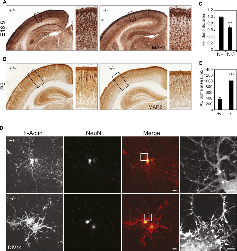
NOMA-GAP regulates dendritic development. (A,B) Loss of NOMA-GAP reduces cortical MAP2 staining. MAP2 staining (brown) in 10-μm coronal sections of littermate E18.5 (A) and P5 (B) brains is shown. Magnifications of the upper cortical layers are shown on the right. Bars: A, 200 μm, 50 μm B, 500 μm, 200 μm. (C) Quantification of the relative dendritic area in the upper layer of P4–P5 animals. n = 5 N+ and 4 N−/− animals; P = 0.0018. (D,E) Loss of NOMA-GAP induces morphological changes and loss of dendritic complexity in mature primary cortical neurons at DIV14. Staining for F-actin (red) and the neuronal marker NeuN (green) is shown for cells derived from littermate N+/− and N−/− E16.5 embryos. Bar, 20 μm. Magnification of the F-actin staining is shown on the right. Bar, 5 μm. (E) The somal area is significantly increased in DIV14 N−/− neurons. n = 21 N+/− and 31 N−/− cells; P = 1.7 × 10⁻⁸.

