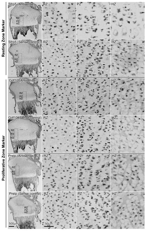
Figure 2. mRNA expression of Sfrp5, Gdf10, and Prelp in growth plate cartilage of 1-week-old rats by in situ hybridization.
Frozen sections were hybridized to 35S -labeled riboprobes. The corresponding sense riboprobe was used as a negative control for each antisense probe. Silver grains were visualized by scanning the slides with ScanScope CS digital scanner (Aperio Technologies, Inc) under bright field microscopy. The left hand panel in each row shows the proximal tibial at low magnification. The other panels show high magnification views of resting zone (RZ), proliferative zone (PZ), and hypertrophic zone (HZ) taken from within the rectanglar area indicated in the corresponding left hand panels. Scale bar, 500μm for low magnification; 50μm for high magnification.

