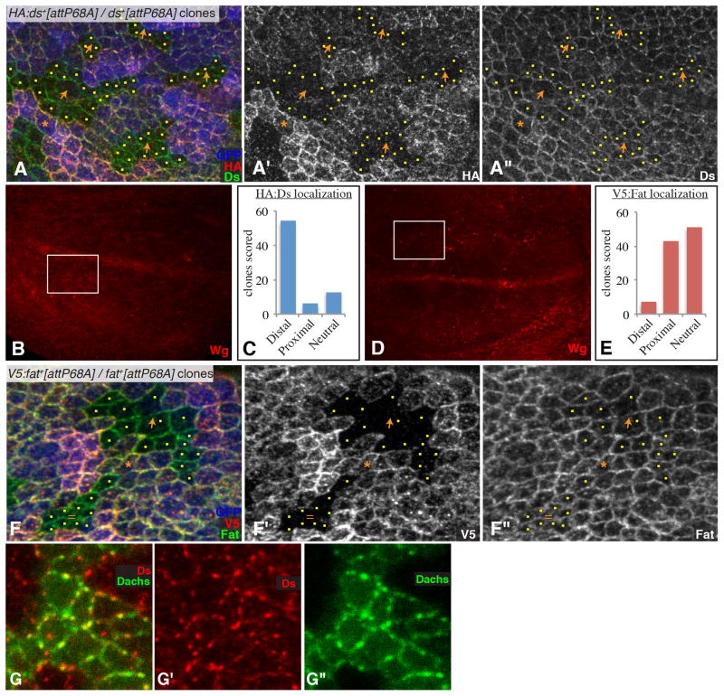
Fig. 3. Polarization of Fat and Ds localization in the wing.
A) Example of a Ubi-GFP attB-P[acman-HA:ds+] FRT80/attB-P[acman-ds+] FRT80 clones in a wing disc, stained for Ds (green/white) and HA (red/white) and labeled by GFP (blue) as indicated. Orange arrows indicate observed polarity of HA:Ds localization, yellow dots mark cells at clone edges. Orange asterisk marks a cell expressing tagged Ds that has unlabeled cells on both its distal and proximal sides; this situation is rare but when it occurs Ds polarization is evident. B) Shows the same wing disc as in (A), but at lower magnification and with Wg expression visible (red), the white rectangle identifies the location of the image in (A). C) Summarizes the results of blind scoring of 73 clones for HA:Ds polarization. D) Shows the same wing disc as in (F), but at lower magnification and with Wg expression visible (red), the white rectangle identifies the location of the image in (F). E) Summarizes the results of blind scoring of 101 clones for V5:Fat polarization. F) Example of a Ubi-GFP attB-P[acman-V5:fat+] FRT80/attB-P[acman-fat+] FRT80 clones in a wing disc, stained for Fat (green/white) and V5 (red/white) and labeled by GFP (blue) as indicated. Orange arrows indicate observed polarity of V5:Fat localization, yellow dots mark cells at clone edges. Equal sign indicates a clone where Fat was scored as not significantly polarized. Orange asterisk marks a cell expressing tagged Fat that has unlabeled cells on both distal and proximal edges, a slight polarization of Fat is evident. G) Close-up of a portion of a wing disc with a Dachs:Cit-expressing clone (green), Stained for expression of Ds (red). Panels marked by prime symbols show individual stains of the image to the left. See also Fig. S2

