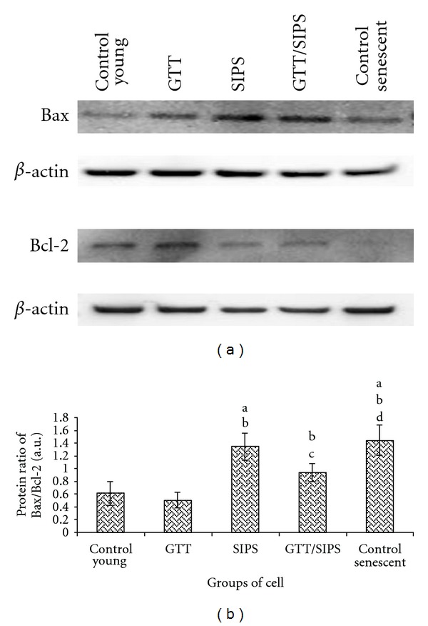
Figure 10.

Representative Western blot of proapoptotic protein (Bax) and antiapoptotic protein (Bcl-2) in HDFs (a). Comparison of Bax/Bcl-2 protein ratio and in different treatment groups (b). Data are expressed as means ± SD, n = 6. adenotes P < 0.05 compared to control young, b P < 0.05 compared to GTT, c P < 0.05 compared to SIPS and d P < 0.05 compared to control senescent cells.
