Figure 2.

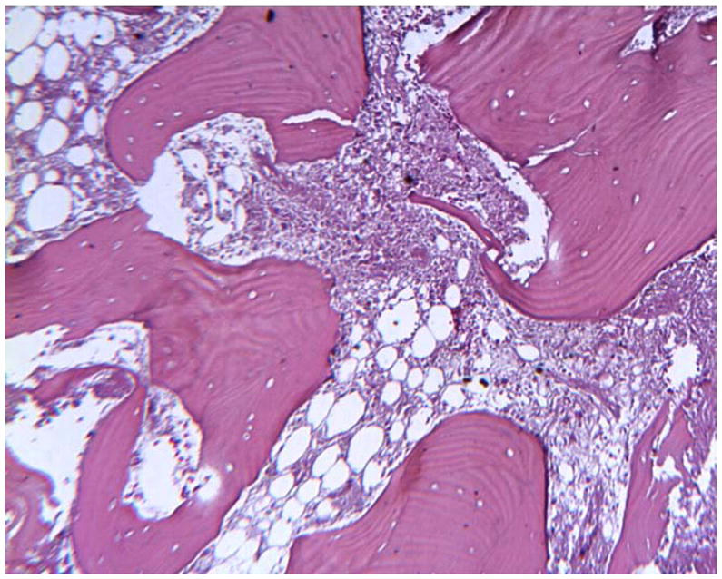
Histology of peri-prosthetic tissues adjacent to a cobalt based TDR showing necrotic bone with empty osteocyte lacunae and necrotic marrow. (H&E, original magnification = 70x). (Color version of figure is available online.)

Histology of peri-prosthetic tissues adjacent to a cobalt based TDR showing necrotic bone with empty osteocyte lacunae and necrotic marrow. (H&E, original magnification = 70x). (Color version of figure is available online.)