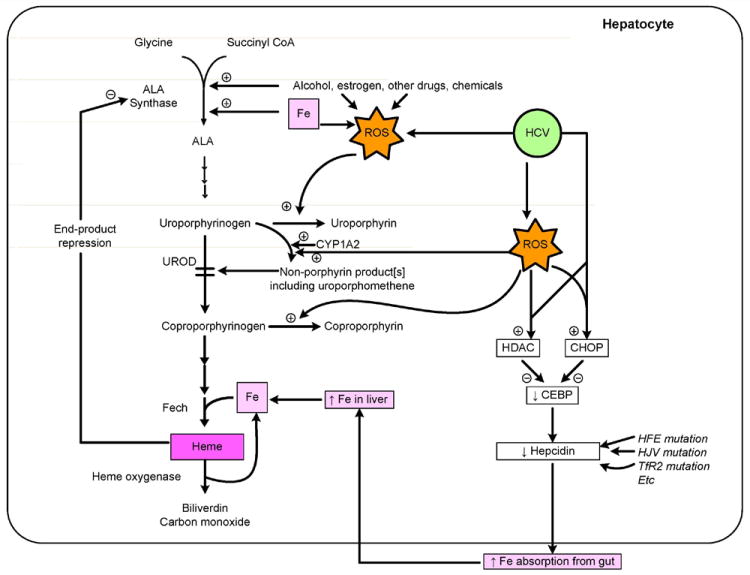
Figure 1. Pathogenesis of Porphyria Cutanea Tarda.

The figure shows a summary of the normal pathway of heme biosynthesis, with emphasis on uroporphyrinogen decarboxylase [UROD] and its inhibition by an oxidation product thought to be derived from uroporphyrinogen. Formation of the inhibitor is thought also to require the action of CYP1A2. Several sites of action of iron are shown, including synergistic induction of ALA synthase 1, increases in oxidative stress (reactive oxygen species [ROS], and induction of heme oxygenase 1. Alcohol and estrogens also increase ROS and induce ALA Synthase 1. HCV infection increases ROS and decreases hepcidin production. The latter decrease is due to up-regulation of histone deacetylase [HDAC] and C/CBP homology protein [CHOP], which result in decreased binding of C/EBP to the hepcidin gene promoter region and thus decreased hepcidin production. Mutations in the HFE gene, [or more rarely, HJV, and/or TfR2 genes] also lead to decreased hepcidin production. The decrease in hepcidin leads to increased absorption of iron from the small intestine, leading to further hepatic iron overload and to amplification of the metabolic disarray.
