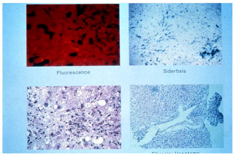
Figure 3. Hepatic Histopathology in PCT.

Fresh unfixed liver fluoresces a bright pin) [upper left, (unstained, 1X] due to excess porphyrins. Typically, there is some degree of iron loading [upper right, (Prussian Blue stain, 10X)] and fatty change and inflammation [lower left, (H&E stain, 20X)]. The latter are often due to alcohol and/or chronic hepatitis C. Sometimes, cirrhosis and/or hepatocellular carcinoma develop [lower right, (H&E stain 10X)]. Photomicrographs kindly provided by JR Bloomer.
