Fig 4.
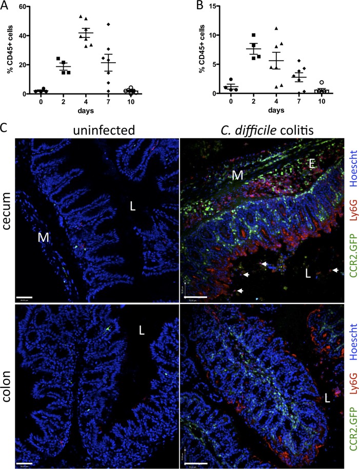
Monocyte and neutrophil infiltration of the cLP in C. difficile-infected mice. C57BL/6 mice were infected with 103 CFU C. difficile following a single dose of clindamycin the day before. Leukocytes were isolated from the cLP and analyzed by flow cytometry to determine neutrophil (A) and monocyte (B) infiltration during the course of infection. Results are expressed as the percentage of CD45+ cells in the cLP and are pooled from two independent experiments. (C) CCR2.GFP mice were challenged with C. difficile following clindamycin administration and sacrificed 3 days later. Frozen sections of cecum and colon were stained with 1A8 antibody (red, anti-Ly6G) to identify neutrophils. Hoechst (blue) was used for nuclear staining. L, lumen; E, edema; M, muscle layer. White arrows point to neutrophils within the luminal content. Scale bars represent 50 μm. Images are representative of 3 independent experiments with 2 to 5 mice each.

