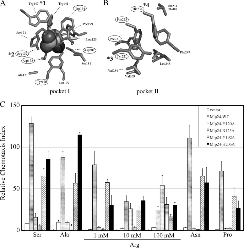
Fig 6.
Mutations introduced into the potential ligand-binding pockets of Mlp24. (A and B) Side chains of the residues constituting the potential binding pockets I (A) and II (B) of Mlp37 (PDB ID, 3C8C). The alanine molecule (shown with a space filling model) in pocket I (A) exists in the crystal. Residues conserved in Mlp24 are circled. Those corresponding to the Mlp24 mutations constructed in this study are marked with asterisks: *1, Y120A; *2, R125A; *3, Y192A; and *4, H205A. (C) Capillary assays of Vmlp201 (Δmlp24 Δmlp37) cells (derived from O395N1) carrying the vector (pAH901, open bars) or the plasmid encoding wild-type or mutant Mlp24 (wild type, hatched bars; Y120A, dotted bars; R125A, checked bars; Y192A, cross-hatched bars; H205A, closed bars). Capillaries were filled with TMN buffers containing each amino acid (1, 10, or 100 mM) shown at the bottom.

