Fig 5.
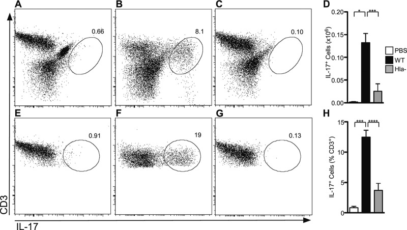
Hla expression is required for induction of a pulmonary Th17 response to S. aureus infection. Flow cytometric analysis of cells stimulated with PMA and ionomycin and then stained with anti-CD3 allophycocyanin (APC) and anti-IL-17 PE. (A to C) Live cell gate from mouse lung following infection with PBS (A), WT S. aureus (B), or Hla− S. aureus (C). (D) Histogram quantifying IL-17+ cells recovered from the lungs of mice. (E to G) CD3+ lymphocyte gate from mouse lung following infection with PBS (E), WT S. aureus (F), or Hla− S. aureus (G). Representative plots are shown. The percentage of IL-17+ cells is indicated. (H) Histogram of the percentage CD3+ cells that are IL-17+. Bars represent the average ± SEM. Results are derived from 4 PBS-treated mice, 21 WT-infected mice, and 14 Hla−-infected mice. P values are indicated: *, P < 0.05; ***, P < 0.001; ****, P < 0.0001.

