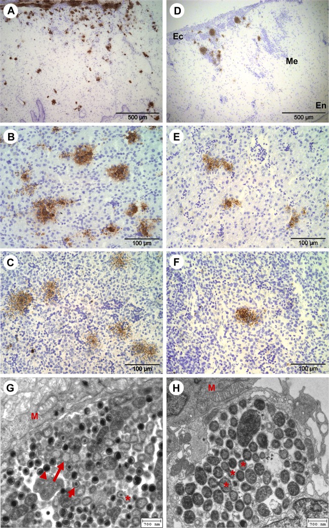
Fig 2.
(A to F) Images of immunohistochemical staining of Chlamydia (brown) and cell nuclei (blue) in CAM and embryonic liver and spleen at 8 dpi. (A) C. psittaci in CAM. (B) C. psittaci in liver. (C) C. psittaci in spleen. (D) C. abortus in CAM (Ec, ectoderm; Me, mesenchyme; En, endoderm). (E) C. abortus in liver. (F) C. abortus in spleen. (G and H) Electron microscopic images of intracellular C. psittaci in CAM of the chicken embryos. Panels G and H show 5 dpi and 6 dpi, respectively. Arrowhead, elementary body; long arrow, reticulate body; short arrow, intermediate body; asterisk, cell division of RBs; M, mitochondria.

