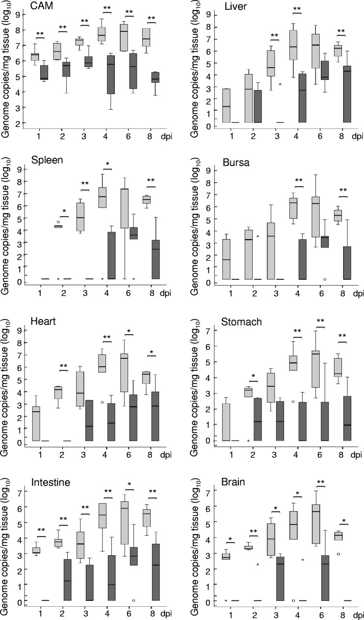
Fig 3.
Quantification by real-time PCR of C. psittaci (light gray bars) and C. abortus (dark gray bars) in CAM and internal organs of challenged embryonated chicken eggs. Chlamydiae were quantified by including decimal dilutions of a genome copy standard. The final load is given in genome copies per 1 mg tissue. Medians are significantly different (*, P ≤ 0.05; **, P ≤ 0.01) between C. psittaci- and C. abortus-infected tissues (calculated by Mann-Whitney U test).

