Fig 1.
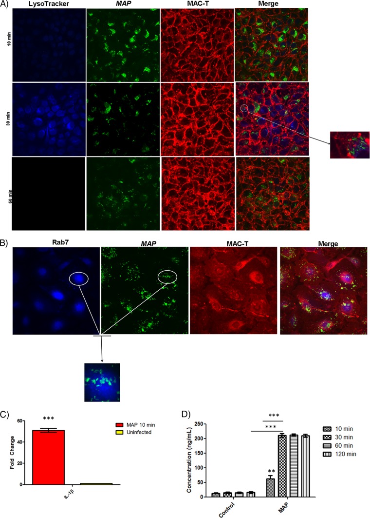
M. avium subsp. paratuberculosis (MAP) induces phagosome acidification and IL-1β processing at the epithelium interface. (A) Confocal microscopy of phagosome acidification in MAC-T cells. MAC-T cells were assessed for phagosome acidification at 10, 30, and 60 min after infection with M. avium subsp. paratuberculosis using LysoTracker blue. Approximately 60% of MAC-T cells contained M. avium subsp. paratuberculosis. M. avium subsp. paratuberculosis induced phagosome acidification at 10 and 30 min postinfection but ceased at 60 min postinfection. (B) Rab7 stain of MAC-T cells infected with M. avium subsp. paratuberculosis 30 min postinfection. LysoTracker staining was validated with indirect staining for the late endosomal marker Rab7. Infected MAC-T cells were positive for Rab7. (C) qRT-PCR of uninfected and infected MAC-T cells at 10 min postinfection. In stark contrast to uninfected cells, M. avium subsp. paratuberculosis-infected MAC-T cells showed a 50-fold upregulation of IL-1β. (D) IL-1β protein levels in uninfected and infected MAC-T cells. Infected MAC-T cells reached peak IL-1β expression at 30 min postinfection. **, P < 0.01; ***, P < 0.001.

