Fig 2.
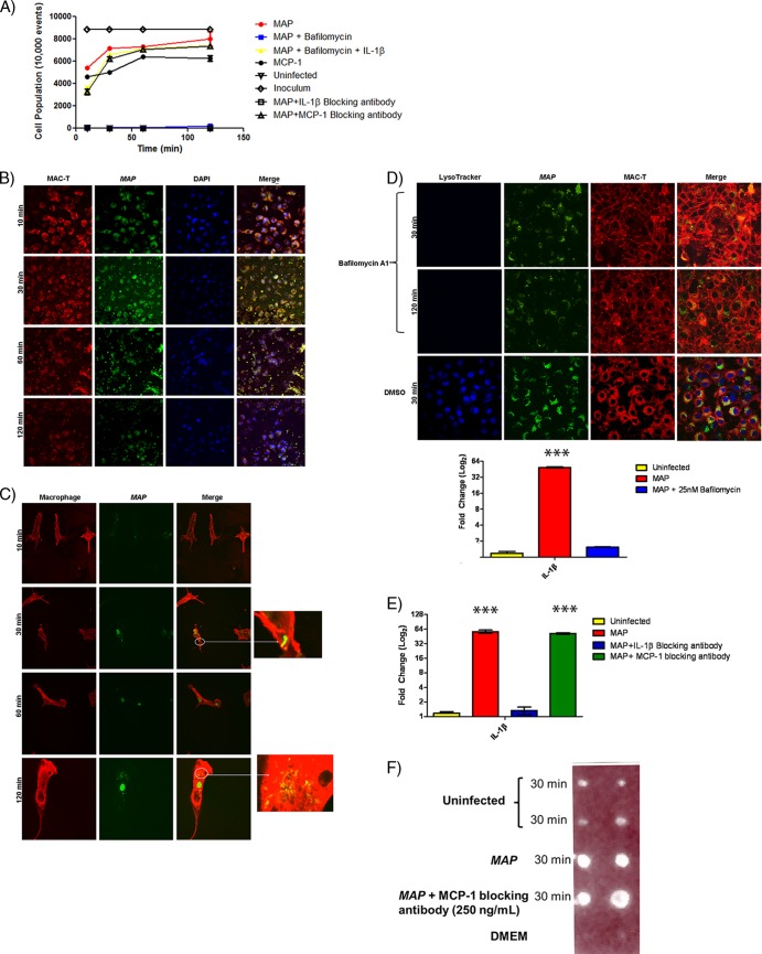
M. avium subsp. paratuberculosis (MAP) enlistment of IL-1β-recruited macrophages to the initial site of infection. (A) FACS analysis of macrophage recruitment in response to M. avium subsp. paratuberculosis infection in the epithelium-bovine macrophage coculture system. M. avium subsp. paratuberculosis infection readily recruits macrophages to the apical chamber. Macrophage recruitment was abolished when phagosome acidification was blocked with bafilomycin A1 or IL-1β expression was prevented. Recruitment was rescued in bafilomycin A1 treatment with the addition of recombinant IL-1β protein. MCP-1 blocking did not impact macrophage recruitment. (B and C) Confocal microscopy of a coculture infection with M. avium subsp. paratuberculosis. (B) Macrophages are readily recruited to the apical chamber, as the Transwell contained only MAC-T cells. (C) Macrophages were found only in the apical chamber. Recruited macrophages contained M. avium subsp. paratuberculosis. (D) Bafilomycin A1 treatment abrogated phagosome acidification and IL-1β transcription (qRT-PCR). Confocal microscopy of bafilomycin A1 treated cells and vehicle control (DMSO) (top). Bafilomycin A1 treatment blocked IL-1β transcription, and results are indistinguishable from those of uninfected controls (bottom). (E) qRT-PCR of infected MAC-T cells treated with IL-1β blocking antibody or MCP-1 blocking antibody. MCP-1 blocking does not impact IL-1β expression. (F) IL-1β dot blot. Cell supernatants were collected during M. avium subsp. paratuberculosis infection of MAC-T cells. The addition of the MCP-1 blocking antibody did not influence IL-1β protein levels. ***, P < 0.001.

