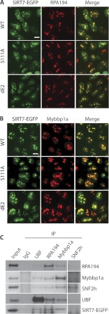
Fig. 5.
Validation of SIRT7 association with Pol I, UBF, and B-WICH components via co-localization and reciprocal isolation. A, co-localization of Pol I (RPA194) with three SIRT7-EGFP fusion proteins. Cellular localization was assessed in HEK293 cells stably expressing the EGFP-tagged SIRT7 variants by immunofluorescence microscopy using anti-RPA194 (red) and anti-GFP (for SIRT7-EGFP, green) antibodies. Bars, 10 μm. B, co-localization of Mybbp1a with SIRT7-EGFP fusion proteins. Immunofluorescence microscopy was performed as in A using anti-Mybbp1a antibodies. C, reciprocal immunoprecipitations (IP) using antibodies against endogenous UBF, RPA194, Mybbp1a, and SNF2h. Equal amount of IgG was conjugated to protein A/G-agarose beads as negative control. Precipitates were analyzed by SDS-PAGE followed by immunoblotting using individual antibodies.

