
Figure 2. Anatomic Reference and Map Nomenclature.
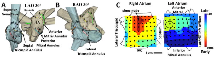
A, B. Electrodes Illustrated Within Patient-Specific Atria (NavX system, St Jude Medical, MN) in 30° LAO and 30° RAO projections, showing alternate splines and electrodes for clarity. C. Sinus Rhythm Map on Biatrial Schematic. Activation at basket electrodes, shown as dots, is displayed as a color-coded map from the sinus node to the lateral inferior LA. The RA is opened between its poles with tricuspid annulus opened laterally and medially; the LA is opened along its equator, with mitral annulus opened superiorly and inferiorly. The pulmonary vein ostia are indicated by dashed lines.