
Figure 4. Localized Electrical Rotors (Spiral Waves) in Human AF, Revealed by Computational Mapping.
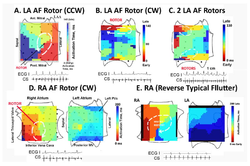
A. Solitary Counterclockwise (CCW) Rotor During Persistent AF in the posterior LA; B. CW Rotor in Persistent AF in the anterior LA; C. Two Concurrent Rotors During Paroxysmal AF in the anterior LA (CW) and inferior LA (CCW); D. Right Atrial Rotor (CW) During Paroxysmal AF in the mid-posterior wall, with fibrillatory conduction to the LA. By Contrast, E. Clockwise Rotor of Reverse Typical Atrial Flutter differs from AF, with 1:1 activation throughout the RA that engages Bachmann’s bundle to activate the LA and no fibrillatory conduction. Key: ECG lead I, CS=coronary sinus electrogram.