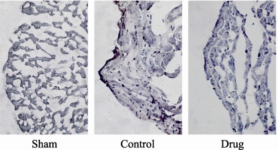
Figure 5. Immunohistochemical evaluation of caspase-3 expression following MI/R injury.

Representative photomicrographs of sections (× 400) from rats subjected to sham myocardial ischemia, ischemia/ reperfusion (30 minutes/2 hours) treated with vehicle, or ischemia/ reperfusion (30 minutes/2 hours) treated with GXST are shown. Dark staining indicates the presence of caspase-3.
