
Fig. 10.
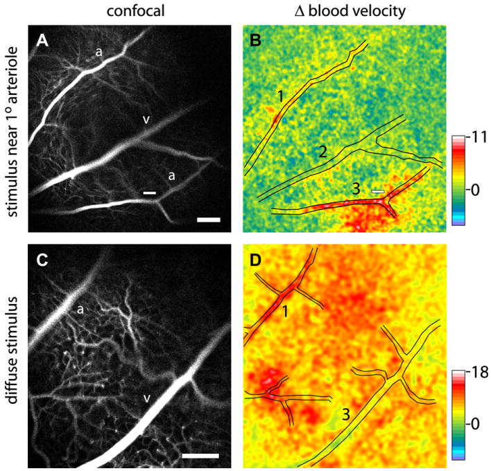
Blood velocity increases evoked by focal (top) and diffuse (bottom) flickering light stimulation in the rat retina. The left panels show confocal images of the retina and right panels show blood velocity changes measured with laser speckle flowmetry. Focal stimulation (small white bars in A and B) evokes a local increase in blood flow (red region in B) while diffuse stimulation evokes a blood flow increase over the entire retina (red and yellow regions in D). Pseudocolor scale bars indicate percent change in blood velocity; from Srienc et al. (2010).