
Fig. 17.
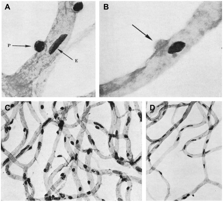
Loss of pericytes in diabetic retinopathy. A, Normal human retinal capillaries showing intramural pericytes (P) and endothelial cells (E). B, Mural ghost pericyte (arrow) in a human diabetic retina. C, Capillary meshwork in a healthy retina, showing a normal ratio of endothelial cells to pericytes. D, Capillary meshwork in a diabetic retina. Note marked loss of pericytes (round nuclei) with preservation of endothelial cells (elongated nuclei). Trypsin digestion, periodic acid-Schiff (PAS) and hematoxylin stain; from Speiser et al. (1968).