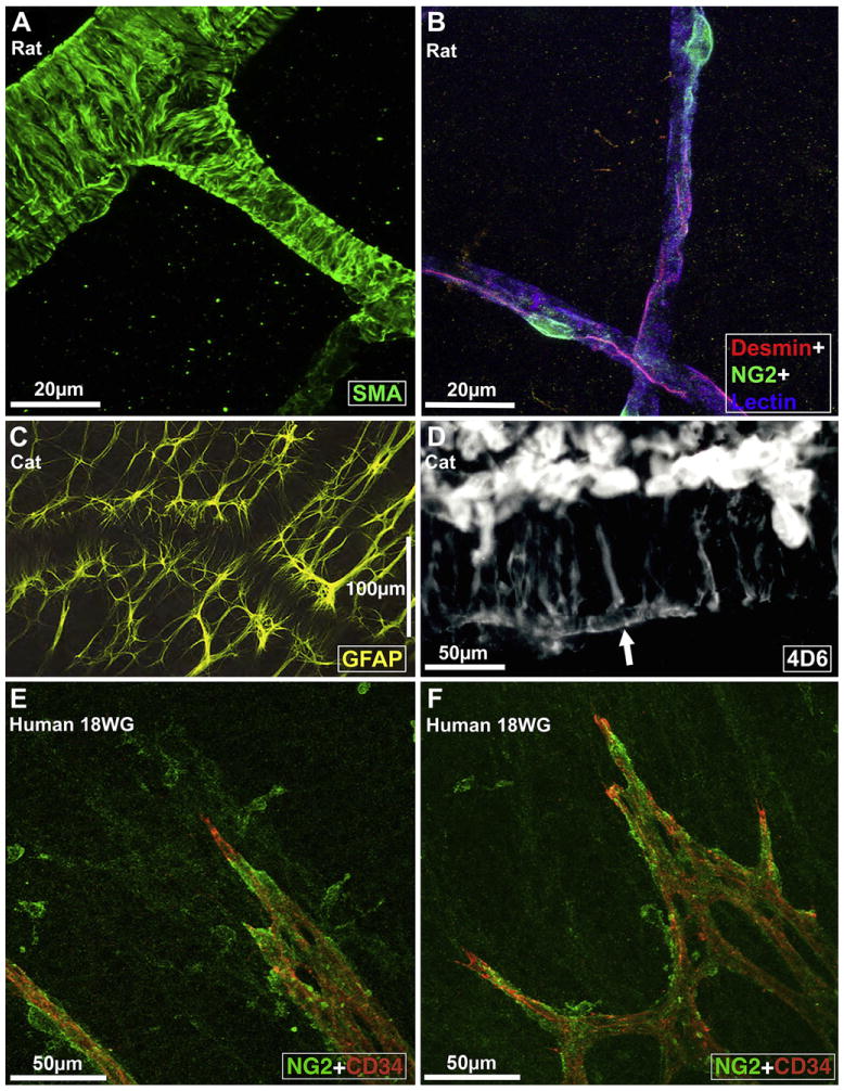
Fig. 3.

Fine structure of retinal vessels and glia ensheathment. A, Rat retinal arterioles (postnatal day 21) labeled for smooth muscle actin (SMA), a smooth muscle cell differentiation marker (from Hughes and Chan-Ling, 2004). B, Capillaries in the adult rat retina triple labeled with Griffonia Simplicifolia isolectin B4 (GS) lectin (blue), anti-desmin (red), and anti-NG2 (green); from Hughes and Chan-Ling (2004). C, Morphology of astrocytes immunolabeled with GFAP (at the level of the superficial vascular plexus) in the cat retina wholemount. Note close association between astrocytes, the vessel wall, and the nerve fiber bundles that run diagonally across the field of view; from (Chan-Ling, unpublished). D, The broken edge of a retinal wholemount, labeled with 4D6, a monoclonal antibody specific to Müller cells; from Dreher et al. (1988). The inner endfeet of Müller cells (top) are brightly fluorescent. At the outer margin of the inner nuclear layer Müller cell processes outline a capillary (arrow); from Dreher et al., 1988. E and F: Micrographs showing the intimate relationship between NG2+ pericytes and the CD34+ angiogenic tip cells of the developing retinal blood vessels in human retinal wholemounts at 18 weeks gestation. The NG2+ pericytes are located just ahead of the leading edge of patent CD34+ vessels; from Chan-Ling et al. (2011b).
