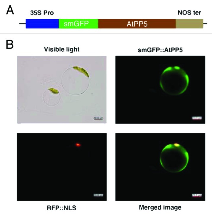
Figure 3. Subcellular localization of AtPP5 fused with GFP in Arabidopsis protoplasts. (A) Schematic diagram of plasmid construct for transforming plant cells for fluorescent confocal microscopy. Expression of the fused genes was driven by the CaMV 35S promoter (35S Pro) and terminated by the nopaline synthetase terminator (NOS ter). Arabidopsis protoplasts were transformed with the resultant constructs and fluorescent images were obtained 12 to 48 h after transformation. Green and red images are GFP and RFP fluorescence signals of smGFP::AtPP5 and RFP::NLS, respectively.
