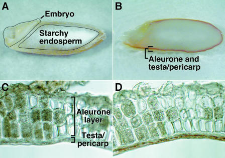
Figure 7.
Proanthocyanidins in H. vulgare Grain Are Concentrated in the Testa.
Imbibed H. vulgare grain were sectioned longitudinally ([A] and [B]), and isolated aleurone layers were sectioned transversely ([C] and [D]). The deep-red peripheral band of vanillin staining in (B) is lacking in the unstained grain (A). The same section through the aleurone layers and attached testa is shown before (C) and after (D) staining. Note that only the region immediately outside of the aleurone layer is stained with vanillin.

