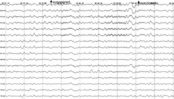
Figure 1.
Electroencephalography pattern from 20-year-old patient with overlapping Bickerstaff’s brainstem encephalitis, Miller Fisher syndrome, and Guillain-Barré syndrome. A diffuse slow wave EEG pattern with 4-6 cycles per second is seen in all electrodes. Twenty-two to twenty-six cycles per second waveform predominates over the centroparietal area, bilaterally. The alpha rhythm over the occipital electrodes was absent. By the time of examination, the patient’s consciousness level demonstrated intermittent drowsiness, suggesting a diagnosis of encephalitis.

