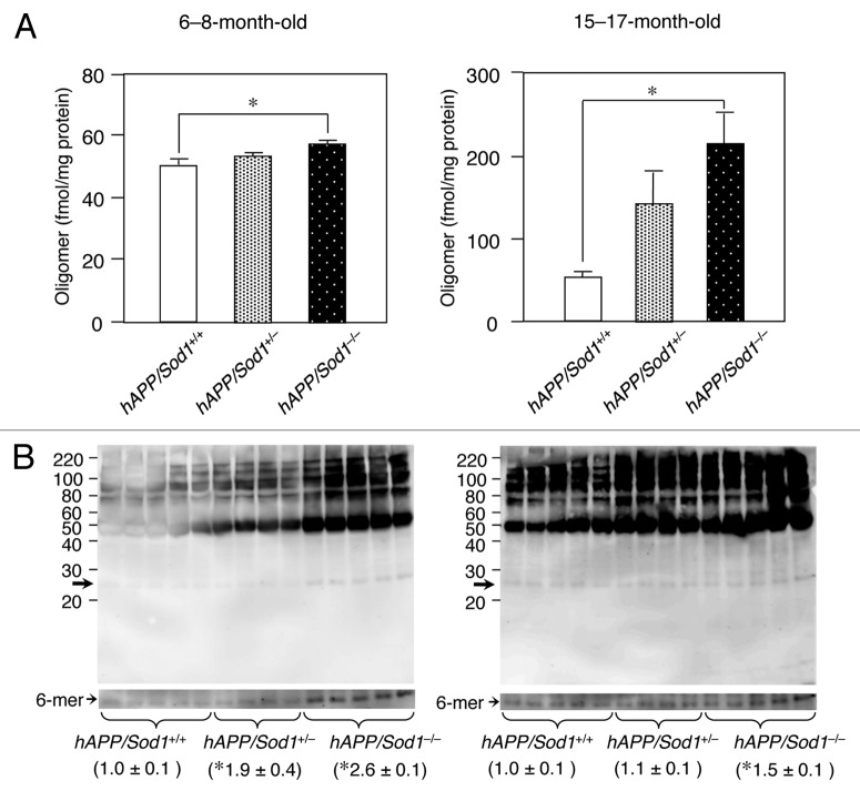
Figure 1. An increase of Aβ oligomer by Sod1 deletion in Alzheimer mice. (A) ELISA analysis of 82E1-specific oligomers using the TBS-soluble fraction of brains of mice (n = 5~7 per genotype) of the indicated genotypes and age. In ELISA for Aβ oligomers (Immuno Biochemical Laboratories: IBL, Gunma, Japan), the same N-terminal Aβ antibody (82E1) is used both for antigen capture and detection. In the younger ages (6−8-mo-old), the level of only Aβ oligomers was significantly increased in hAPP/Sod1-/- as compared with the hAPP/Sod1+/+ mice, but not Aβ42 or Aβ40 (ref. 15). On the other hand, older (15−17-mo-old) hAPP/Sod1-/- showed a significant elevation of Aβ oligomers as well as Aβ42 and Aβ40 (ref. 15) as compared with the hAPP/Sod1+/+ mice. These data are rearrangement of the previous work (ref. 15). (B) Distribution of Aβ aggregates by western blotting of mice (n = 4~5 per genotype) of the indicated genotypes and age. The detailed procedure was described previously (ref. 15). In brief, Tris buffered saline (TBS)-soluble fractions (2 µg/µL) were subjected to western blotting using 10–20% Tricine gel (Invitrogen) and transferred to a PVDF membrane (0.2 μm pore size, Bio-rad). Anti-Aβ antibody (6E10, 1:1,000, Signet) was used for Aβ detection. Overexposed bands corresponding to the hexamer (arrows: ~30 kDa) were used for relative quantification. left: 6−8-mo-old, right: 15−17-mo-old. *p < 0.05 vs. hAPP/Sod1+/+, mean ± s.e.m.

An official website of the United States government
Here's how you know
Official websites use .gov
A
.gov website belongs to an official
government organization in the United States.
Secure .gov websites use HTTPS
A lock (
) or https:// means you've safely
connected to the .gov website. Share sensitive
information only on official, secure websites.
