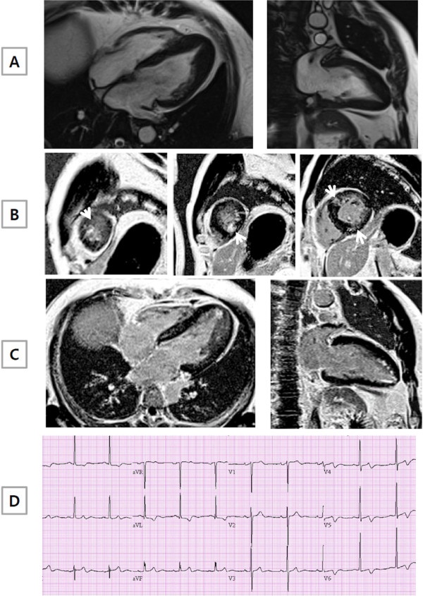
Figure 2.
A representative example of ApHCM with late gadolinium enhancement (LGE) by CMR. (A) Long axis CMR cine images clearly demonstrated the presence of apical hypertrophy, as in Figure 1. In contrast, short (B) and long axis (C) CMR images showed diffuse LGE at the basal and mid interventricular septum or RV insertion site (non-hypertrophic segment) and at the apex (hypertrophic segment), as indicated by the arrow. (D) Negative T wave inversion in anterior leads of electrocardiogram was present.

