Fig. 3.
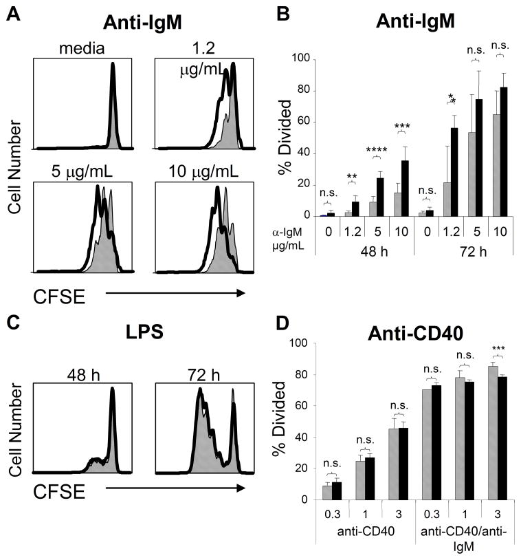
gp91phox KO B cells display enhanced BCR-induced proliferation. Purified WT (grey bars) or gp91phox KO (black bars) B cells were loaded with CFSE and stimulated with graded doses of anti-mouse IgM (A, B), LPS (C), or anti-CD40 (D). Proliferation was measured by CFSE dilution at 48 or 72 h. ****p<0.006, ***p<0.02, **p<0.03, *p< 0.04, n.s., not significant; Student’s t-test. Data mean + SD (n≥3 spleens) and are representative of four independent experiments.

