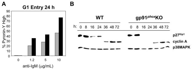
Fig. 4.
More gp91phox KO B cells enter the cell cycle after BCR ligation. (A) 24 hr after stimulation via the BCR, WT and gp91phox KO B cells were incubated with 2 μg/mL Pyronin Y. Pyronin Y staining was measured by flow cytometry. WT (grey bars) and gp91phox KO (black bars). (B) Purified WT and gp91phox KO B cells were stimulated with 5 μg/mL anti-IgM F(ab′)2. Cells were cultured for the indicated time periods, collected and lysed. Western blots of cell lysates with anti-p27Kip1 or anti-cyclin A. All data are representative of three independent experiments.

