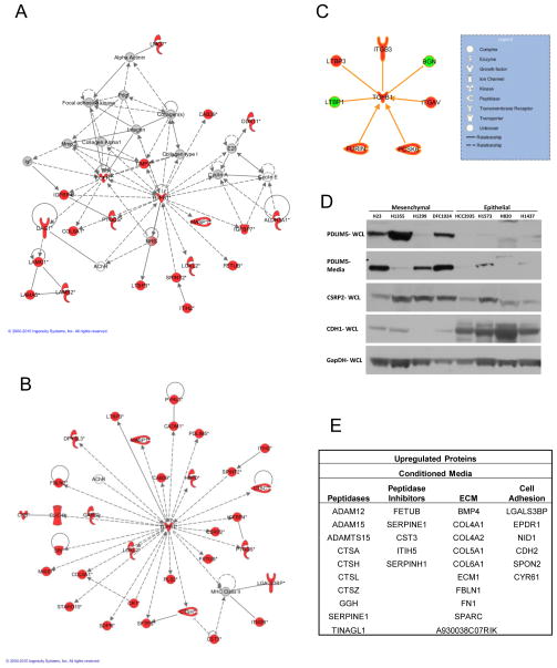
Figure 3. TGFβ signaling in metastatic cells.
A. The most significant network from Ingenuity Pathway Analysis for upregulated proteins in the conditioned media shows evidence of TGFβ-1 regulation. B. Network analysis of upregulated proteins combined from conditioned media, cell surface and total cell extracts reveals a stronger and more significant node than from the individual compartments C. Proteins directly interacting with and regulating TGFβ-1. Network objects colored red indicate upregulation and green objects indicate downregulation. D. Western blots comparing protein expression in epithelial and mesenchymal NSCLC cell lines. E. Microenvironment related proteins downregulated with miR-200 expression.

