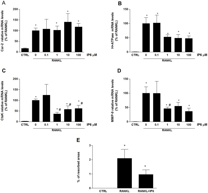
Figure 3. IP6 directly inhibits RANKL-induced osteoclast bone resorption ability.
RAW 264.7 cells were treated with RANKL (100 ng/ml) for the generation of OCL and IP6 for 5 days and gene expression of osteoclast functional markers was determined: Car-2(A), H+-ATPase (B), CtsK (C) and Mmp-9 (D). Data represent fold changes of target genes normalized with GAPDH mRNA and 18s rRNA, expressed as a percentage of RANKL-dosed cells non-treated with IP6, which were set to 100%. Values represent the mean ± SEM. Significant differences were assessed by Student’s t test: *p≤0.05 versus control cells. #p≤0.05 versus RANKL treated cells. (n = 6) (E) Bone resorption ability of RAW 264.7 cell treated with 1 µM of IP6 during osteoclastogenesis was evaluated by resorption pit assay on dentine discs (n = 3). Data represent the percentage of the resorbed area by osteoclasts. Values represent the mean ± SEM. Significant differences were assessed by Mann-Whitney test:*p≤0.05 versus untreated cells. #p≤0.05 versus RANKL treated cells.

