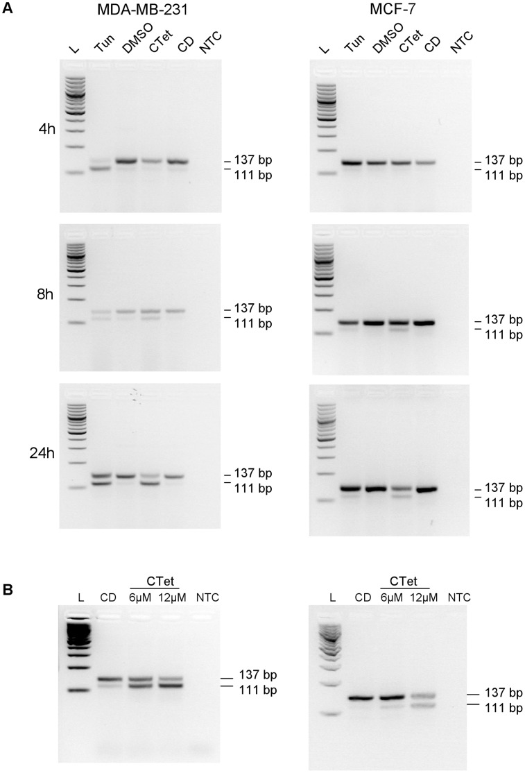
Figure 3. Alternative splicing of Xbp-1 mRNA in CTet-treated cells.
MDA-MB-231 (left) and MCF-7 cells (right) were treated with CTet or tunicamycin, and total RNA was reverse transcribed and analyzed by PCR for detection of alternative spliced forms, as described in methods. (A) Cells were treated with 12 µM CTet or 2 µg/ml tunicamycin for 4, 8 and 24 h. Cells treated with γ-cyclodextrin and DMSO were used as negative controls for CTet and tunicamycin treatment, respectively. (B) Cells were treated with 6 µM and 12 µM CTet for 24 h. Cells treated with γ-cyclodextrin were used as control. One representative experiment is shown for each cell line. L, 100 bp DNA ladder; Tun, tunicamycin; CD, γ-cyclodextrin; NTC, no template control.

