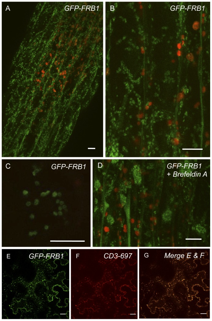
Figure 5. GFP-FRB1 fusion protein accumulates in subcellular compartments.
A. Hypocotyl cells of transgenic plants expressing GFP-FRB1 fusion protein under the control of a constitutive promoter. Note that the GFP accumulates in subcellular compartments (green). Red fluorescence is from chloroplasts. B. GFP-FRB1 fluorescent particles in the cortical cytoplasm. C. Higher magnification image showing the ring morphology of the GFP-tagged compartments. D. Hypocotyl cells of GFP-FRB1 plants treated with 100 µg/ml Brefeldin A for 15 min. Note the redistribution of the GFP-labeled compartments to aggregates, especially around nuclei. Some of the GFP signal has also become soluble. E. Transient over-expression of GFP-FRB1 fusion protein or F. mCherry (CD3–967) fusion protein in an epidermal tobacco cell. G. Micrograph showing overlap of E and F. Scale bars equal 10 µm in A–D and 20 µm in E–G

