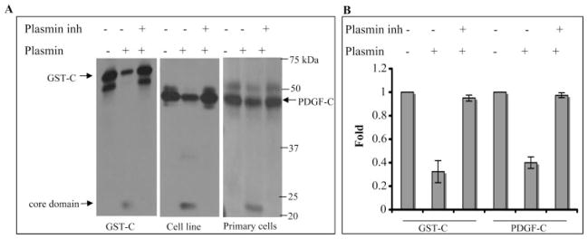
Figure 2.
Plasmin processed PDGF-C. (A) Plasmin (0.2 μg) was preincubated with or without α2-plasmin inhibitor (2 μg) for 1 hour at room temperature and was added to GST-PDGF-C (GST-C, 60 ng, left) or conditioned medium (40 μL) from either an RPE cell line (middle) or primary fetal RPE cells (right). After 10-minute incubation at 37°C, the samples were subjected to PDGF-C Western blot analysis. Top bands: latent PDGF-C is the higher molecular mass in the right-hand panel (GST adds 26 kDa), and the core domain is the lower molecular mass species. (B) Quantification of the Western blot results from three independent experiments (mean ± SD). GST-PDGF-C (68% ± 9%) and latent PDGF-C (60% ± 5%) were processed by plasmin. The plasmin inhibitor blocked the processing of GST-C and native PDGF-C by 95% ± 3% and 97% ± 2%, respectively.

