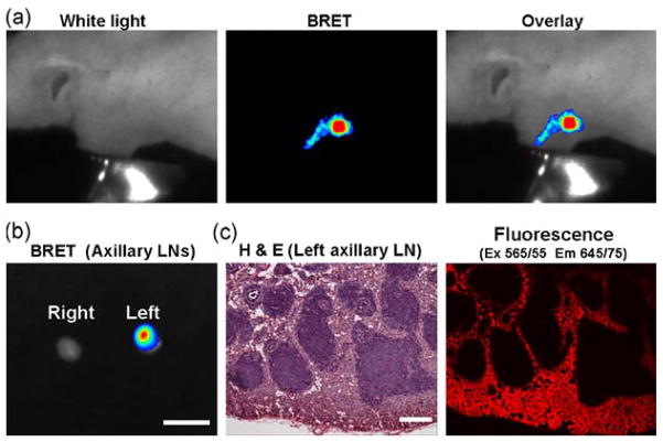
Figure 2.
BRET lymphatic image of the mouse receiving BRET-Qdot655 injection at the left paw. In vivo image demonstrates lymphatic duct and lymph node (a). Accumulation of BRET-Qdot655 in lymph node was confirmed by ex vivo imaging (b) and histological analysis (c). Bars on ex vivo image and H&E image are 5 mm and 100 μm, respectively.

