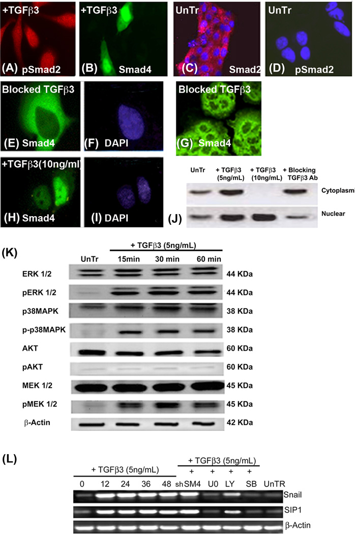
Fig. 5. TGFβ3 induces Smad-dependent and Smad-independent pathways in the palatal seam cells to activate the EMT related transcription factors, Snail and SIP1.
Immunofluorescence was performed on homogenous palatal MES cells that were treated TGFβ3 (5 and 10ng/mL) for 3 h to demonstrate the Smad translocation in response to TGFβ3. In the presence of 5ng/mL of TGFβ3, pSmad2 and Smad4 are seen in the cytoplasm and nucleus (A and B respectively). In the absence of TGFβ3, Smad2 (C) remains in the cytoplasm while pSmad2 (D) is not seen in either nuclei (DAPI) or cytoplasm. MES cells treated with blocking TGFβ3 antibody for 24 h showed no expression of Smad4 in the nucleus (E), but only in the cytoplasm (F, DAPI), and there was no change of morphology in MES cells as they remained epithelial (G). Once the dose of exogenous TGFβ3 was increased to 10ng/mL for 3 h, Smad4 completely moved to nucleus (I, DAPI), and no cytoplasmic expression was observed (H). Immunoblotting analysis of nuclear and cytoplasmic proteins from untreated MES cells (control) and those treated with TGFβ3 (5ng/mL), demonstrated that Smad4 protein expression was higher in both the cytoplasm and nucleus compared to its low expression in control cells. However, cells treated with 10ng/mL TGFβ3 showed only nuclear expression of Smad4. MES cells treated with blocking TGFβ3 antibody showed much higher expression of cytoplasmic Smad4 protein than nuclear (Fig. J). (K) MES cells were treated with TGFβ3 (5ng/mL) for 15, 30, and 60 min., and protein from these cells was extracted for immunoblotting – control cells were untreated (UnTr) at 0 (not shown) and 60 min (shown). Results showed phosphorylation of ERK, p38MAPK and MEK1/2, but not AKT in MES cells treated with 5ng/mL exogenous TGFβ3. These proteins were not phosphorylated in control, untreated cells harvested at either 0 (not shown) or 60 min. (L) mRNA was collected from the MES cells treated with TGFβ3 (5ng/mL) for 0, 12, 24, 36, and 48 h. Some groups of MES cells were treated with a Smad inhibitor (Smad4 shRNA; shSM4) for 48 h and Smad-independent pathways were blocked using small synthetic chemical inhibitors – LY294002 to block PI3 Kinase (20µM), SB202190 for p38MAPK (20µM) and U0126 for MEK1/2 (20µM) – for 60 min. mRNA expression of Snail and SIP1 transcription factors increased dramatically within 12 h of TGFβ3 treatment and remained high until 48 hours. When Smad4 was inhibited, mRNA expression of these transcription factors stayed unaffected compared to 48 h of TGFβ3 treatment. However, Snail and SIP1 mRNA expression was drastically reduced when cells were treated with inhibitors of the Smad-independent pathways, except for PI3 kinase.

