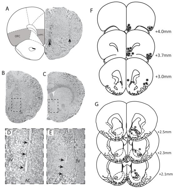
Figure 1.
Representative brain sections demonstrating the localization of stimulating electrode placements (arrows) within the mPFC and OFC (A) and recording electrode locations within the lateral (B expanded in D) and medial (C expanded in E) NAc. The locations of each identified recording electrode (f: lateral octagons - medial squares), stimulating electrode (G: star) or infusion site (g: circle) are depicted with reference to a stereotaxic atlas adapted from Paxinos and Watson (Paxinos and Watson, 1986).

