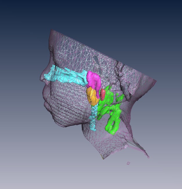
Figure 2.

Surface rendering of the head and neck and three-dimensional reconstruction of the upper airway and lymphoid tissues of the subject shown in Figure 1: airway (light blue), adenoid (magenta), tonsils (orange), retropharyngeal nodes (red), and deep cervical lymph nodes (green).
