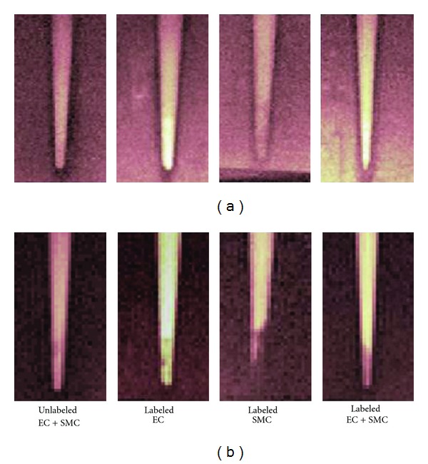
Figure 6.

(a) T 1-weighted and (b) T 2-weighted MR images of labeled cell pellets at 7 Tesla. EC were labeled with Gd2O3. SMC were labeled with SPIO. Imaging was performed at 1 day post-labeling. Scan parameters were T R/T E = 1000/7.4 ms for T 1-weighted scans, T R/T E = 4000/41.9 ms for T 2-weighted scans.
